Turku PET Centre – One of the Best in the World
Turku PET Centre produces an exceptionally wide range of radiopharmaceuticals on a worldwide scale. In a unique way, the centre can both examine individual patients’ diseases and advance medical research.
The national Turku PET Centre is jointly run by the University of Turku, Åbo Akademi and the Hospital District of Southwest Finland and is situated right at the heart of the Turku University Hospital. The centre bustles with activity as five PET scanners constantly scan either patients or volunteers for research.
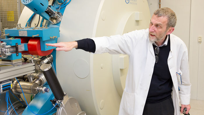
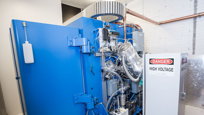
In the basement of the building, behind a two-meter thick concrete doors are the centre of the whole operation: the particle accelerators.
The first particle accelerator of the PET Centre was acquired in 1974, reveals Professor of Radiochemistry Olof Solin, who works at the PET Centre.
– We mainly produce Carbon-11, Fluorine-18, Oxygen-15 and Copper-64, adds Solin.
The diversity of radionuclides means that a wide range of radiopharmaceuticals can be produced in the radiochemistry laboratory which can then be used for instance in diagnosis of various diseases.
– We have a wide range of different radiopharmaceuticals, possibly more than anywhere else in the world, says Professor of Radio Chemistry Olof Solin. The core of the pharmaceuticals is the radionuclides which are created with a particle accelerator.
Elevated Glucose Metabolism Reveals Cancer
Olof Solin has experienced the development and growth of the PET Centre. When Åbo Akademi acquired its first particle accelerator in 1974, Solin was writing his Master’s thesis and chose radionuclide production as his topic.
– We started researching short-lived radionuclides which are suitable for imaging, Solin reminisces back for nearly 40 years and then describes shortly in a nutshell what happens in positron emission tomography.
– The radionuclides produced with the particle accelerator decay due to positron emission. Positrons are the antiparticles of electrons, antimatter that cannot exist in our world. When a positron is generated, it immediately starts looking for an electron to interact with. Then the mass transforms into pure energy, into two gamma rays which are emitted in opposite directions, Solin describes the process.
The first particle accelerator was acquired for the research of nuclear physics but it was clear from the beginning that its capacity would be used for medical purposes as well. The researchers built the first imaging scanners themselves, the first actual PET scanner was acquired in 1988.
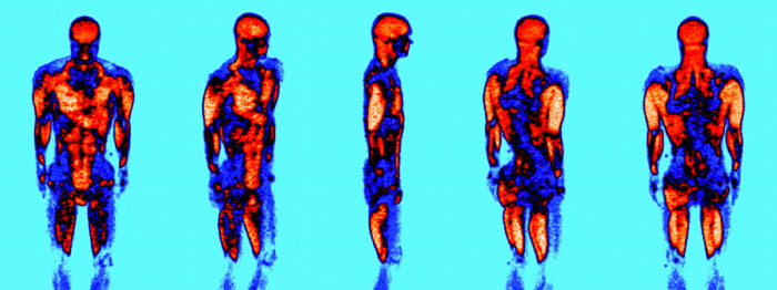
Solin tells how the PET scanner can recognise the decay process and define the trajectory of the quanta. With this information, the scanner can produce a precise image on, for example, where in the body the glucose uptake is high and where it is low.
– For example, cancer tissue uses glucose as a fuel. When we have added Fluorine-18 to glucose and injected the tracer into the patient, we can see with the scanner in which part of the body the glucose uptake is elevated. The cancer tissue can be seen in the image as a high accumulation of the tracer, says Solin.
Even Seconds Count in Different Stages of the Process
Only a few meters from the particle accelerator is the radiopharmaceutical chemistry laboratory and as a part of that the cleanrooms which can only be entered by the trained personnel.
– A radionuclide has to be combined with a molecule so that it forms a radiopharmaceutical. The molecule controls how the radiopharmaceutical, called tracer, is localised in the body. Before injection, each batch of radiopharmaceutical has to undergo a rigid quality control to ensure the safe use of the radiopharmaceutical, Solin emphasizes.
As the radionuclides and radiopharmaceuticals are short lived, each minute and, with some radiopharmaceuticals, even seconds are important.
– The half-life – i.e. the time it takes for half of the nuclides to decay – of the used radionuclides ranges from two minutes to two hours. That’s why we have to produce the nuclides ourselves, Solin tells.
PET imaging requires radioactive isotopes to be administered to the patients or subjects being imaged, the radiotracers have a short half-life which for example enables multitracer studies, several tracers can be injected into the patient during the same day enabling sophisticated study protocols.
At the PET Centre, researchers constantly develop new radiopharmaceuticals and methods of combining radionuclides with molecules. At the moment, for example, researchers are trying to find suitable radiopharmaceuticals for tracing the treatment of Alzheimer’s disease and diabetes.
Half of the Capacity Used for Patients, the Other Half for Research
On the upper floors there are five PET scanners. On a normal week day, each one of them is in constant use. About half of the capacity is used for diagnostics of individual patients and the other half is reserved for research.
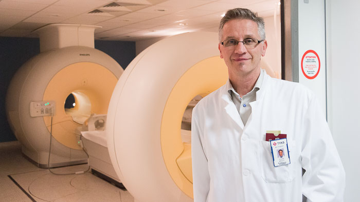
– The original idea was to use 30 percent of the capacity for diagnostic imaging of patients, but that share has risen to about 50 percent. This is a national centre and we have responsibility to help patients from all around the country. It is the right thing to do and we are happy that we can provide clinical applications as well, says Juhani Knuuti, the director of Turku PET Centre.
PET imaging is used all around the world mainly to diagnose cancer.
– In addition to cancer, we study the brain, heart, metabolism and inflammations. In research, the variety is even more extensive, says Knuuti.
The radioactivity is one of the big challenges in the work. Therefore, automatised synthesis methods have been developed for the laboratories.
The applications are a result of devoted development work. The researchers of the PET Centre have managed to develop several new radiopharmaceuticals that can be used to examine diseases that could not be detected with PET scanners before.
The basic funding comes from the universities and the hospital district and rest of the funding consists of academic research grants from the Academy of Finland and the EU. Part of the operations of the PET Centre are funded by industrial cooperation.
Investment in Facility and New Devices Fixes the Bottleneck at Turku PET Centre
The centre that started out with only a couple of employees now employs about 150 people.
– All the parties – University of Turku, Åbo Akademi University and the Turku University Hospital – have understood how valuable the PET Centre is and are ready to invest in it. We have also had very good directors, says Olof Solin.
The investment can be seen in the amount and quality of research.
– In the last few years, about 6 to 8 dissertations have been published each year and over a hundred peer-reviewed publications have been published, most of them in the top scientific journals of the world, says Solin.
Turku PET Centre is taking a giant leap into a new era. The Centre has purchased a new cyclotron and now it can produce radioactive isotopes much faster than before. Also, a new faster and more sensitive PET/CT-camera is installed.
Turku is establishing a top position in positron emission tomography (PET) on a worldwide scale. The next step is to expand the Centre’s facilities to serve the increased production of radioactive isotopes. The owners of the PET Centre as well as the Academy of Finland, that has granted funding, are responsible for the investment.
– Seven new lead shielded hoods, hot cells, have been installed to a new 100 m2 clean room area next to the existing clean rooms. This allows us to double our capacity to produce various radiopharmaceuticals in a modern environment, says Juhani Knuuti.